21st Eibsee Meeting
Author: Lis de Weerd, PhD student in the lab of Prof. Christian Haass and Hans and Ilse Breuer Foundation stipend holder
What makes our brain cells break down when a person develops dementia? And what’s going on inside these cells when that happens? These questions are the main topic of discussion at the Eibsee Meeting: a yearly conference organized by Prof. Christian Haass (DZNE and LMU, Munich), which brings together renowned scientist in the field of neurodegeneration to share and discuss their latest research findings.
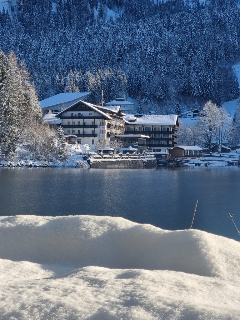
The 21st edition of the Eibsee meeting took place this year from December 6th to 8th at the Eibsee Hotel in Grainau, located at the beautiful Eibsee lake and surrounded by the Bavarian Alps. The heavy snow of the weekend prior made it a challenge to reach the destination by train, but thanks to the hard work of the organization committee all invited guests made it in time for a warm welcome by Prof. Haass in the afternoon to kick off the program.
The Eibsee meeting presents a diverse program, with an informal scientific walk around the lake, keynote lectures, session talks, flash talks and a poster session. A new feature this year were the two young investigator keynote lectures, giving new group leaders the chance to present their current work and future plans. Below I will share with you some of the highlights of this year’s meeting.
Hitchhiking inside a neuron
Hitchhiking is a way of traveling by getting rides from people who are going in the same direction as you by chance, making it a cost-efficient way to travel. The cells in our body have evolved to function as cost-efficiently as possible, so it may be no surprise that hitchhiking also happens inside a brain cell. Prof. Michael Ward from The National Institutes of Health (NIH) in Bethesda, USA, gave the first keynote lecture on how molecules can hitchhike in neurons to their final destination.
More specifically, Prof. Ward explained how RNA, a molecule that helps translate genetic instructions from DNA to build proteins in our cells, hitchhikes down the long nerve ends called axons, where it is translated to makes proteins. Rather than jumping into its own ‘car’ (using motor proteins that use up a lot of energy), RNA molecules hitch a ride with lysosomes that are traveling down the axon. Lysosomes are also known as the cell’s recycling centers, where proteins are broken down and their building blocks (amino acids) can be used to build new proteins. So, are these recycled amino acids used by RNA that hitched a ride to make new proteins in the axon? Indeed, using clever microscopy techniques, Prof. Ward showed that this was the case. The understanding of this process can help to understand how the loss of functional lysosomes can lead to frontotemporal dementia (FTD) and amyotrophic lateral sclerosis (ALS).
The role of supporting cells: microglia & brain vasculature
Neurons are not the only brain cells that are involved in neurodegenerative diseases. This was highlighted by the two invited young investigator keynote speakers: Dr. Xianyuan Xiang from the Chinese Academy of Sciences in Shenzhen, China and Dr. Andrew Yang from the University of California (UCSF) in San Francisco, USA.
Xianyuan started her own group in 2021 after completing her PhD and short post-doc in the lab of Prof. Christian Haass, where she investigated the role of the risk gene TREM2 in Alzheimer’s disease. TREM2 is a gene expressed in microglia, the main immune cell type in the brain. Microglia support neurons by responding to injuries or infections and cleaning up debris such as dead cells. During her keynote lecture Dr. Xiang presented the ways in which she continues to investigate the multifaceted role of TREM2 and microglia in her new lab, not only in the context of Alzheimer’s disease, but also in cancer and stroke.
Dr. Andrew Yang is a self-proclaimed engineer-turned-neuroscientist, who started his group in 2022, after a post-doc investigating the brain vasculature in the lab of Prof. Tony Wyss-Corey. He likened the brain vasculature to a home of the brain, which has a thermostat to control temperature, a door for which you need a specific key and means of communicating to the rest of the world such as Wi-Fi. All of these functions are also performed by the blood vessels in the brain, which controls brain temperature, forms a selective barrier that only allows certain molecules to pass through and carries molecules from other parts of the body to the brain. And if the home is damaged it might not be enough to fix the interior (the neurons), but also the exterior (the blood vessels). Dr. Yang presented in his keynote lecture how he investigates all these functions of the brain vasculature using multiple discovery approaches to identify genetic changes in aging and neurodegenerative diseases.
Crossing barriers
The second keynote lecture of the meeting was given by Dr. Gilbert di Paolo from Denali Therapeutics Inc., located in South San Francisco, USA. At Denali, Dr. di Paolo leads a team of scientists that have developed strategies to shuttle proteins into the brain (using a modified key that can open the door in the blood vessels). He showed the therapeutic application of this strategy in the context of FTD, where in specific cases the main cause is the lack of a protein called progranulin (PGRN). Using the modified key (PTV) attached to PGRN (PTV:PGRN), it can enter the brain and rescue all phenotypes observed in models. The drug is now in clinical trials. Dr. di Paolo’s talk showed not only a successful crossing of barriers between the blood vessels and brain, but also between academic and industry research.
Scooters and protein folds
Neurodegenerative diseases are often characterized by the aggregation of sticky protein in the brain. Certain diseases such as ALS and FTD can share the same sticky protein clumps but present with a very different disease. How is that possible? To answer this question the final keynote speaker of the conference, Dr. Benjamin Ryskeldi-Falcon, looks at these proteins under a microscope at the highest resolution possible: at the atom level. To achieve this, his lab extracts proteins from post-mortem human brains and places them under a microscope that bombards the proteins with electrons. From the way the electrons bounce off the protein, Dr. Ryskeldi-Falcon can visualize the protein structure. And what he sees? Surprisingly, each neurodegenerative disease has proteins that are folded in different ways, giving them a unique structure: one looks like a scooter, whereas another resembles a chevron badge. By knowing how these structures are different, we can use this information to develop drug that prevent these specific folds.
In summary, the 21st Eibsee meeting provided a fantastic opportunity for scientific exchange, bringing together early career researchers and established investigators. The unique setting allows for many networking opportunities, resulting in the formation of many new and exciting scientific collaborations. It is with many thanks to the invaluable support of the Hans and Ilse Breuer Foundation, SyNergy and DZNE, which make this conference possible.